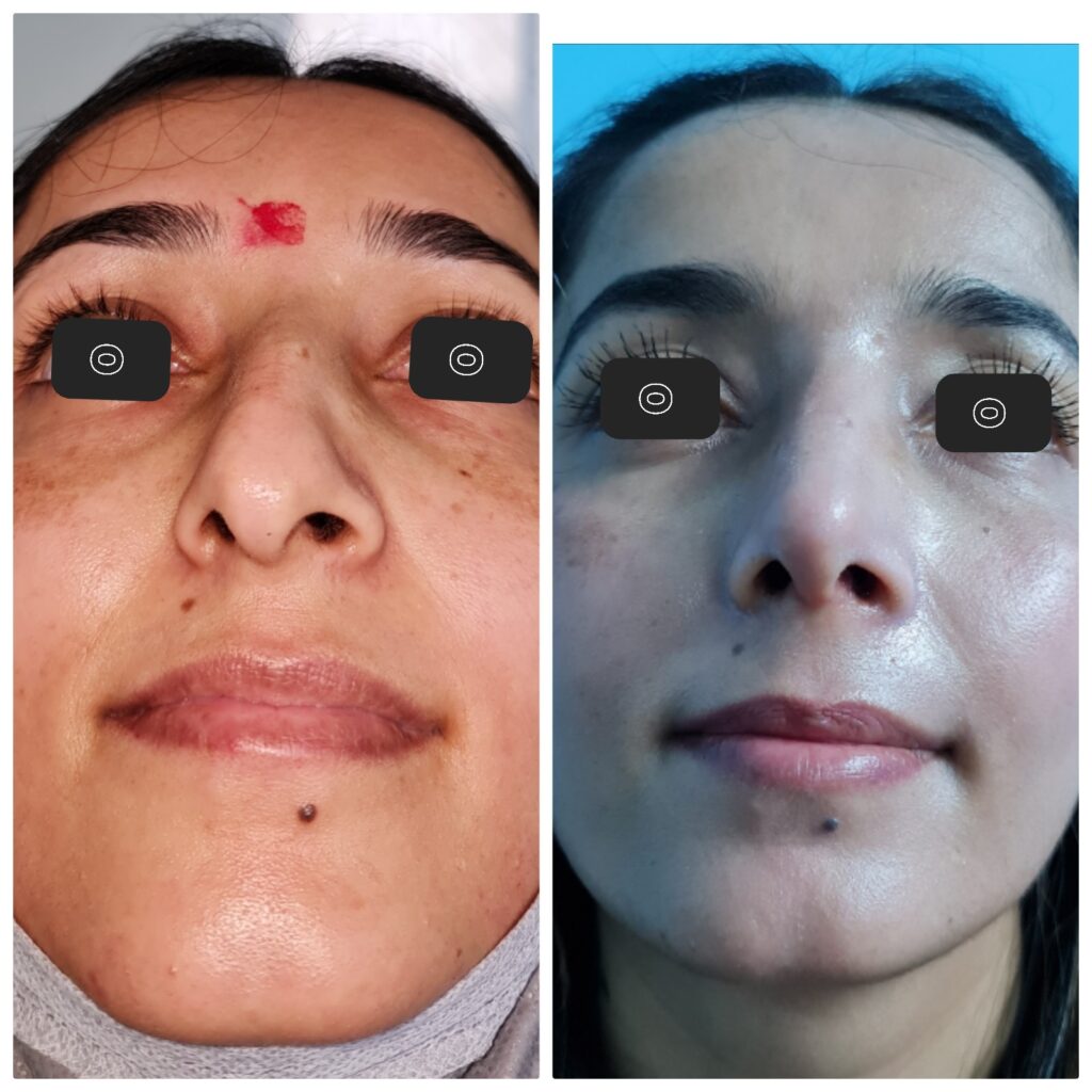
Septoplasty for Functional problem

Nasal septoplasty is one of the most commonly performed procedures by an ENT surgeon. The primary indication for this functional surgery is usually septal deviation resulting in significant and symptomatic nasal airway obstruction. Many surgical techniques and approaches have been described; these include endonasal, endoscopic and open procedures. Septoplasty can also be performed alongside or in addition to rhinoplasty, turbinoplasty, or as part of functional endoscopic sinus surgery to improve surgical exposure and access. Other indications for septoplasty include recurrent epistaxis, obstructive sleep apnea, sinusitis, and facial pain and/or headaches due to septal spurs that contact a turbinate (Sluder’s neura). Septoplasty may also be necessary in conjunction with endoscopic sinus, skull base, or orbital surgery in order to provide better access to target structures.
Technique
The patient is positioned supine with the head tilted slightly towards the surgeon. Prominent nasal hairs are trimmed. Local anesthetic is infiltrated bilaterally in the sub mucoperichondrial plane, using 1% lidocaine with epinephrine (1:100,000) until the mucosa is well blanched. This assists in the hydro-dissection of the planes in addition to analgesia and hemostasis. An incision is made using a #15 blade along this caudal edge to expose the cartilage. Typically, a hemi-transfixion (vertical incision through one side of the membranous septum) or Killian’s incision (vertical incision more posteriorly, over the quadrangular cartilage) is used. If the septoplasty is performed in conjunction with an open rhinoplasty, the septum may be approached from above after the upper lateral cartilage is disarticulated from the dorsal quadrangular cartilage. Dissecting scissors and Freer or Cottle elevators are then used to develop a sub-mucoperichondrial plane and dissect posteriorly to reveal the quadrangular cartilage, PPE, and vomer. Care must be taken not to perforate the mucosa, especially if dissection over bony spurs or deviations is necessary. A second flap on the contralateral side may then be raised through the same incision. If a mucosal perforation has occurred on one side, extreme care must be taken not to perforate the contralateral side; bilateral, opposing perforations can lead to a septal perforation post-operatively. As the surgeon reaches farther posteriorly, longer nasal specula will be required to obtain good visualization.
Evaluation of bony and cartilaginous deformity is made. Various instruments can be used to incise the septum, and sometimes these are used in combination. A blade or a Freer elevator are used to remove the deviated piece of cartilage. To maintain nasal dorsum and tip stability, an “L-strut” shape of quadrangular cartilage is preserved (dorsal and caudal edges); the posterior aspect of the cartilage is, therefore, often the area that is removed. In the event that the deviation involves the L-strut, an extracorporeal septoplasty or an open approach with grafting may be required. The dorsal and caudal limbs of the L-strut, as well as their attachments to bone, should remain 10 to 15 mm in width to guarantee sufficient support for the external nose. A twisting movement is often employed to remove sections of cartilage or bone; care must be taken not to twist too forcefully outside of the anterior-posterior axis as this can cause a fracture of the cribriform plate and potential leak of cerebrospinal fluid.
The mucoperichondrial flaps are laid back into position against the septum. Interrupted stitches using absorbable suture material (e.g., chromic gut) are used to close the incision. Running mattress, or “quilting,” sutures are often made through and through the septum to close any dead space and reapproximate the flaps to avoid hematoma accumulation post-operatively. If the mucoperichondrial flaps remain completely intact at the end of the operation, some surgeons will place a small incision inferiorly in one flap to facilitate the egress of any fluid that may collect within the septum. Some surgeons prefer to use a septal stapler rather than placing a quilting suture by hand. Care should also be taken to avoid disruption of the keystone area during the removal of deviated portions of the septum.
Removed cartilage should be saved in normal saline or a surgical gauze sponge dampened with normal saline. Pieces of native cartilage can be reshaped and used to reinforce the L-strut if necessary or to contribute to grafts used in rhinoplasty.
The mucoperichondrial flaps are laid back into position against the septum. Interrupted stitches using absorbable suture material (e.g., Vicryl) are used to close the incision.
Running mattress, or “quilting,” sutures are often made through and through the septum to close any dead space and reapproximate the flaps to avoid hematoma accumulation post-operatively. If the mucoperichondrial flaps remain completely intact at the end of the operation, some surgeons will place a small incision inferiorly in one flap to facilitate the egress of any fluid that may collect within the septum.An antibiotic cream can be applied intranasally.